Variables
Independent:
1. Source of anti-fungal compound
Dependent:
1) Presence/ absence of zone of inhibition (around the wells in the PD agar where is seed extract (test) and the ethanol (control) is placed) [Anti-fungal test 1]
2) Diameter of fungal colony [Anti-fungal test 2]
Controlled:
1) Temperature of the growth of fungus (30°C)
2) Concentration of extract (1g in 5ml of 50% ethanol/ Phosphate Buffer Saline)
3) Growth medium (Potato dextrose)
Apparatus & Materials
Apparatus:
1. Micro pipette
2. Pipette filler (Battery-operated)
3. Centrifuge
4. Shaking incubator
5. Mortar and pestle
1. Micro pipette
2. Pipette filler (Battery-operated)
3. Centrifuge
4. Shaking incubator
5. Mortar and pestle
Materials:
1. Extracts
2. Fungus (Aspergillus niger)
3. Ethanol
4. Phosphate Buffered Saline
5. Potato dextrose medium
6. 0.45m micro filter
7. Centrifuge tubes
8. Sterile toothpicks
9. Sterile spreaders
1. Extracts
2. Fungus (Aspergillus niger)
3. Ethanol
4. Phosphate Buffered Saline
5. Potato dextrose medium
6. 0.45m micro filter
7. Centrifuge tubes
8. Sterile toothpicks
9. Sterile spreaders
Procedure
Growth of fungus :
Potato Dextrose Agar (PDA) was prepared and poured in agar plates. The plates were allowed to set overnight. 50ml of PD broth was prepared. Aspergillus niger was inoculated into 10ml of PD broth and was incubated at 30°C overnight.
Preparation of extracts:
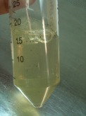
Extracts were prepared in ethanol (Capsicum, chili, spring onion bulb and spring onion leaf) or Phosphate Buffered Saline (Barley) with the concentration of 5g in 10ml (50%) using the mortar and pestle. The extracts were centrifuged at 7000 rpm (rounds per minute) and micro filtered to obtain a clear solution.
Anti-fungal test (1):
Wells were punched in the PD agar and were filled with 70µl of extract or control.The extract and control were left to diffuse into the PD agar for an hour. 100µl of fungus was added to 7ml of soft agar and was mixed well. The soft agar was poured over the PD agar as a thin overlay and the plate was incubated at room temperature overnight.
Anti-fungal test (2a):
100µl of extract or control was spread on the PD agar. Aspergillus niger was spot inoculated on the center of the plate using a sterile toothpick. The plates were incubated at room temperature.
Anti-fungal test (2b):
100µl of fungal spores was added with 100µl of extract or control. 100µl of extract or control was spread on the PD agar. Aspergillus niger was spot inoculated on the center of the plate using a sterile toothpick. The plates were incubated at room temperature.
Potato Dextrose Agar (PDA) was prepared and poured in agar plates. The plates were allowed to set overnight. 50ml of PD broth was prepared. Aspergillus niger was inoculated into 10ml of PD broth and was incubated at 30°C overnight.
Preparation of extracts:
Extracts were prepared in ethanol (Capsicum, chili, spring onion bulb and spring onion leaf) or Phosphate Buffered Saline (Barley) with the concentration of 5g in 10ml (50%) using the mortar and pestle. The extracts were centrifuged at 7000 rpm (rounds per minute) and micro filtered to obtain a clear solution.
Anti-fungal test (1):
Wells were punched in the PD agar and were filled with 70µl of extract or control.The extract and control were left to diffuse into the PD agar for an hour. 100µl of fungus was added to 7ml of soft agar and was mixed well. The soft agar was poured over the PD agar as a thin overlay and the plate was incubated at room temperature overnight.
Anti-fungal test (2a):
100µl of extract or control was spread on the PD agar. Aspergillus niger was spot inoculated on the center of the plate using a sterile toothpick. The plates were incubated at room temperature.
Anti-fungal test (2b):
100µl of fungal spores was added with 100µl of extract or control. 100µl of extract or control was spread on the PD agar. Aspergillus niger was spot inoculated on the center of the plate using a sterile toothpick. The plates were incubated at room temperature.
Microfiltered Fungus
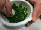
Preparing of Extracts using mortar and pestle
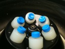
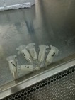
Extracts are centrifuged at 7000 rpm
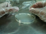
Wells are punched into the PD agar plates
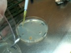
Extracts are added into the wells and left to diffuse into the Agar for one hour
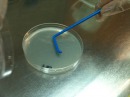
100µl of extract or control is spread on the PD agar.
A. Niger is spot inoculated on the center of the plate using a sterile toothpick
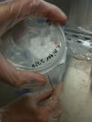
100µl of fungal spores is added with 100µl of extract or control
Data Analysis
After about 2-3 days, the diameter of the fungal colony is checked.
